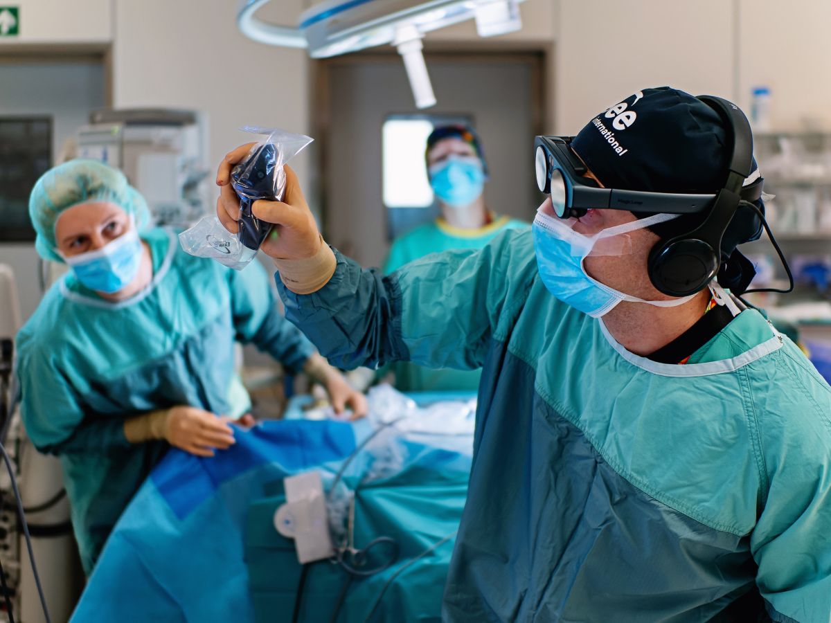
Neurosurgery requires exceptional precision and deep understanding of complex anatomy, often under high-risk conditions. Surgeons must integrate vast amounts of imaging and procedural knowledge to plan and execute safe interventions. Immersive technologies, such as spatial imaging and virtual reality, offer tools for detailed, three-dimensional visualizations of neuroanatomy to support spatial understanding, enhance planning, and improve training in this challenging field.
Important notice regarding surgical planning and professional clinical use
Specialized functionalities for surgical planning and pre-operative professional applications are exclusive to Medical Imaging XR PRO FDA. This version is not yet available. Information about the release date will be published here soon.
Medicalholodeck is currently undergoing the required FDA (U.S. Food and Drug Administration) and CE (Conformité Européenne) certification processes. Our team is working diligently to ensure full compliance with all regulatory standards, and we expect Medical Imaging XR PRO to be available in both the United States and the European Union soon.
For updates on product releases, regulatory progress, and availability, or for any related inquiries, please contact info@medicalholodeck.com.
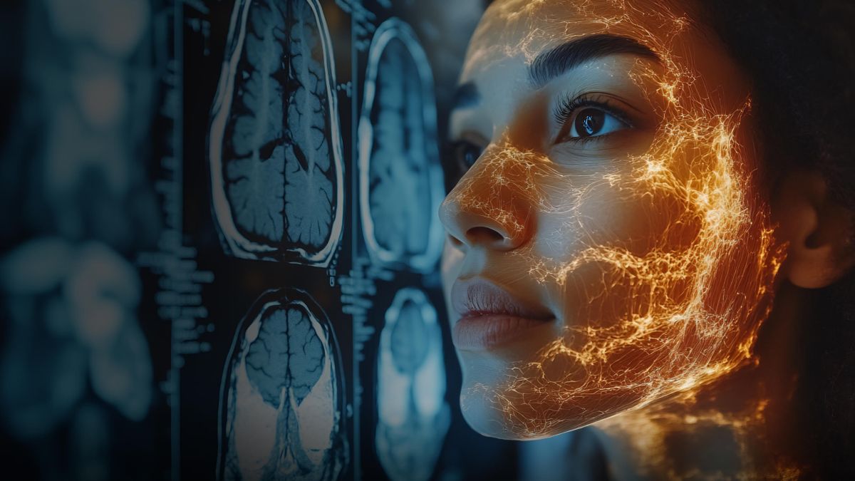
From 2D to 3D: Enhanced anatomical insight
Virtual reality platforms, such as Medicalholodeck, enable neurosurgeons to convert a patient’s CT or MRI data into 3D models and visualize them in immersive environments. These models not only preserve all critical information from 2D imaging but can also be rotated, zoomed, or sectioned in the virtual space. This facilitates the anatomical understanding of neurological structures and tackles the persisting issue of 2D scans – requiring mental reconstructions of the complex anatomy.
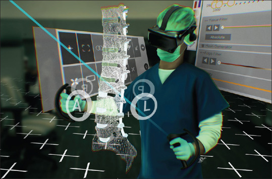
An illustrative rendering depicting a surgeon exploring virtual reality to create a perioperative plan. Of note, is the feasibility to access a perioperative planning environment in an office space. The instrumentation extends from T9-Pelvis
https://doi.org/10.4103/jcvjs.jcvjs_48_23
A surgical team used Medicalholodeck to analyze the spatial relationships of the patient’s spine and its surrounding structures. By exploring different angles the team was able to identify the safest surgical corridors and anticipate potential obstacles. This additional layer of preoperative insight ultimately strengthened confidence in approach selection and supported more precise execution during spinal revision surgery.
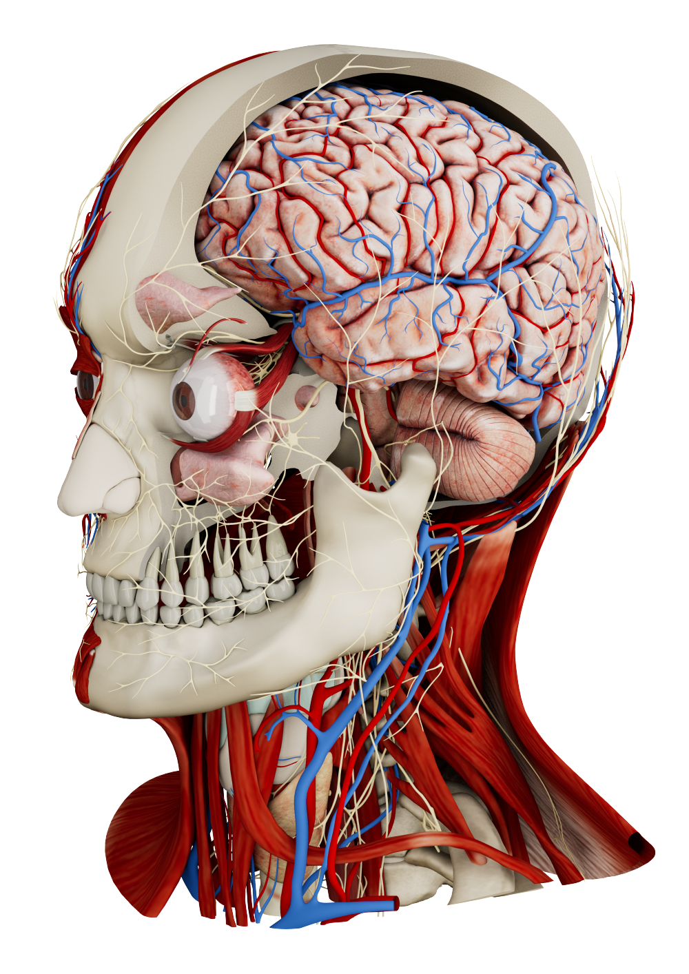
Cranial holograms
Similarly to VR, AR can support surgical planning, but its ability to overlay virtual data onto the real environment makes it particularly valuable intraoperatively. This technology allowed neurosurgeons to plan the optimal approach for resection of a vestibular schwannoma, a benign but challenging tumor due to its proximity to critical neurovascular structures. During the procedure, 3D cranial models were projected onto the patient’s anatomy, enhancing spatial awareness of these critical structures. This visualization confirmed the chosen surgical approach, verified incision sites, and helped optimize patient positioning while accounting for adjacent vessels.
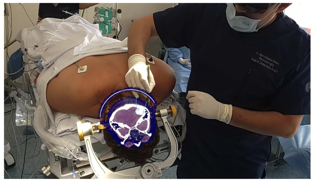
Target placement with AR, where the patient’s CT scan can be observed in the coronal view.
https://doi.org/10.3390/brainsci14101025
AR can also be applied in brain tumors, such as meningioma and ependymoma. In three neurosurgeries, the surgeons benefited from the 3D patient-specific overlays. This real-time guidance enabled more accurate tumor localization, facilitated safer dissection, and improved confidence in surgical execution, contributing to minimal complications and favorable postoperative outcomes.
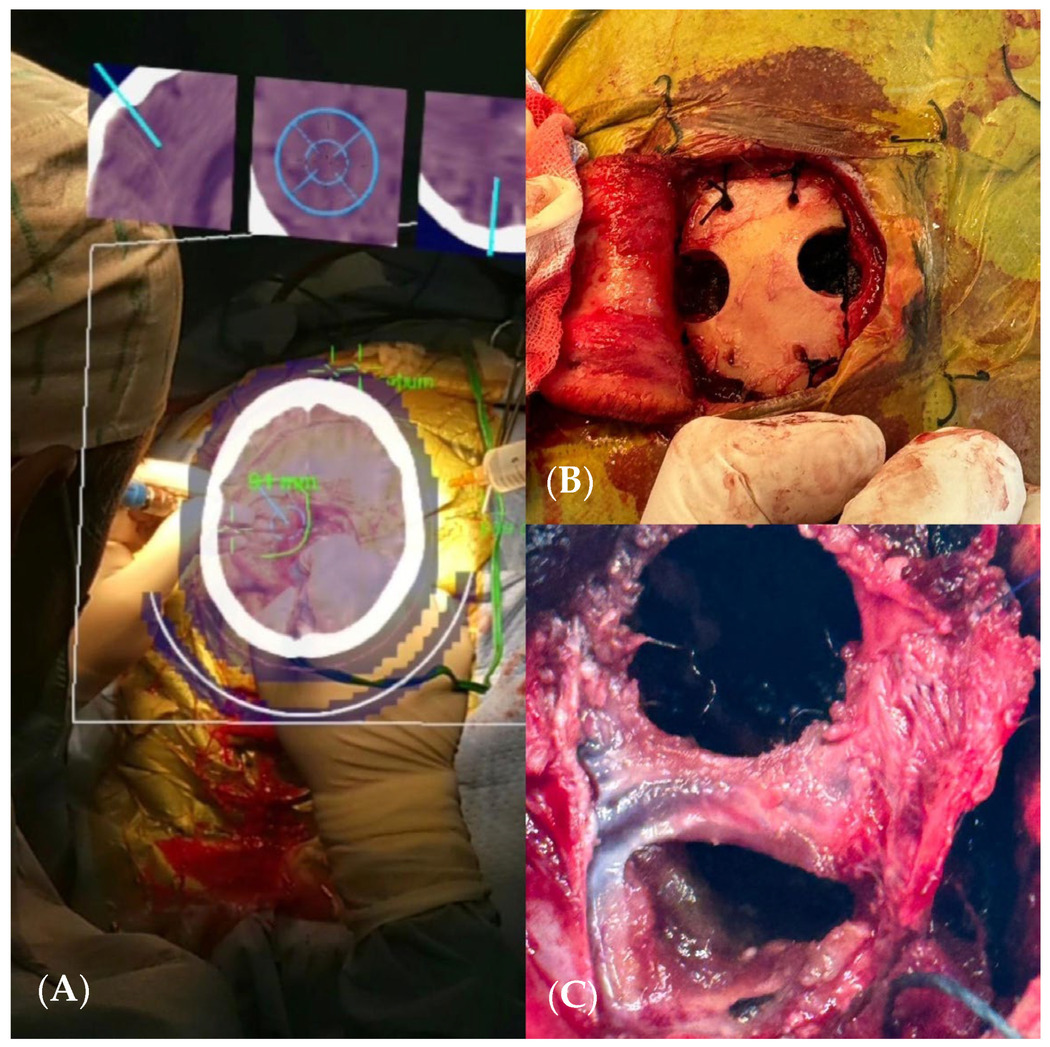
(A) AR view showing a real-time CT scan. (B) Craniectomy assisted by augmented reality. (C) Total resection of the lesion.
https://doi.org/10.3390/brainsci14101025
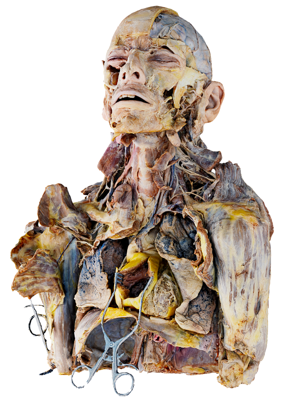
Immersive-to-tactile simulation
Surgical training sessions in VR or AR are increasingly integrated into medical curricula because they enhance skill transfer and procedural confidence. To further improve practice and trainee engagement, a workshop combined an AR mobile application with 3D-printed anatomical models. In the first phase, residents selected neurosurgical approaches and rehearsed each step with AR guidance on the 3D models. In the second phase, they performed the same procedures on physical 3D skull simulators, with each step evaluated by senior neurosurgeons. The residents reported that this hybrid training deepened their understanding of neuroanatomy and improved technical skills, underscoring its potential as a standard component of neurosurgical education.
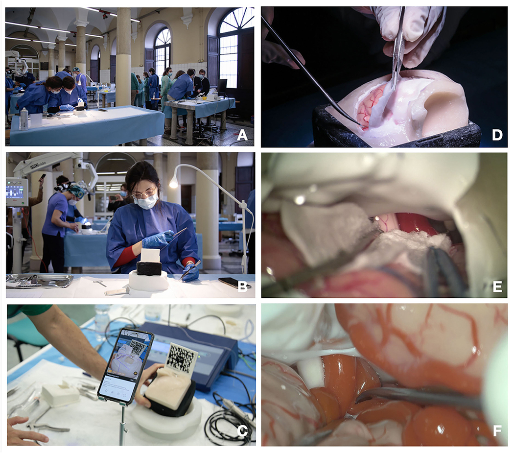
(A–E) Training steps. (A) Training laboratory set up; (B) suturing exercise; (C) augmented reality (AR) for craniotomy and approach planning; (D) dura opening on the pterional approach simulator box; (C) microscopic intradural phase through pterional approach with gentle brain retraction; (E,F) microscopic exploration and dissection through pterional approach of the carotid artery, optic nerve, and sylvian vessels.
https://doi.org/10.3389/fsurg.2022.862948
Patient education
The complexity of many neurosurgical conditions may be difficult for patients to fully grasp, which can lead to misunderstandings during clinician–patient communication. Virtual reality addresses this problem by providing interactive 3D environments that allow patients to explore their own anatomy with accurate spatial context. When combined with the surgeon’s guidance, this approach enhances patients’ understanding of their diagnosis, increases confidence in the planned procedures, and strengthens the informed consent process.
Immersive technologies offer enhanced 3D visualization and spatial understanding, benefiting neurosurgeons in planning and guiding various procedures. They also support medical trainees in acquiring technical skills and help patients better understand their conditions. Overall, these tools are advancing surgical precision, improving outcomes, and enriching education throughout neurosurgical practice.
How to get started
Medicalholodeck integrates with secure hospital systems, providing PACS access, HIPAA-compliant data handling, and full patient data security. It runs on stereoscopic 3D displays, VR headsets, mobile devices, and standard Windows systems, enabling flexible use in hospitals, classrooms, and training centers.
Specialized features for surgical planning are exclusive to Medical Imaging XR PRO. Currently, Medicalholodeck is available only for educational use. The platform is undergoing FDA and CE certification, and we expect Medical Imaging XR PRO to be available soon in the U.S. and EU markets.
For updates, regulatory news, availability, or questions contact info@medicalholodeck.com.