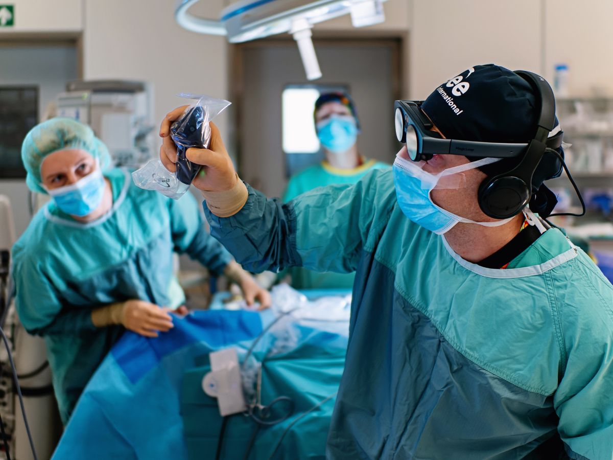
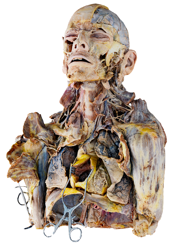
Children present unique medical challenges due to small size, anatomical variability, and the rarity of diseases, which can make diagnosis, surgical planning, and intraoperative decision-making particularly demanding. Spatial imaging, virtual and augmented reality provide powerful tools to facilitate comprehension of complex anatomy, enabling more precise planning and execution of procedures. These technologies have the potential to reduce intraoperative risks and improve outcomes across a range of pediatric surgical specialties.
Important notice regarding surgical planning and professional clinical use
Specialized functionalities for surgical planning and pre-operative professional applications are exclusive to Medical Imaging XR PRO FDA. This version is not yet available. Information about the release date will be published here soon.
Medicalholodeck is currently undergoing the required FDA (U.S. Food and Drug Administration) and CE (Conformité Européenne) certification processes. Our team is working diligently to ensure full compliance with all regulatory standards, and we expect Medical Imaging XR PRO to be available in both the United States and the European Union soon.
For updates on product releases, regulatory progress, and availability, or for any related inquiries, please contact info@medicalholodeck.com.
Precision surgery planning in pediatrics
Yang W, Xu Y, Wang Z, Ye M, Chen R, Da M, Qi J (2025) Virtual reality-assisted preoperative planning for pediatric thoracoscopic segmentectomy: a retrospective study. BMC Pediatrics.
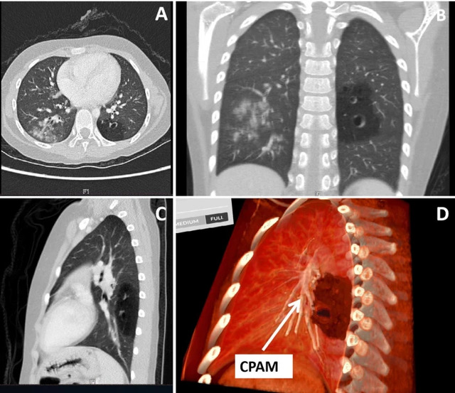
The report describes how pediatric surgeons used Medicalholodeck's virtual reality medical imaging to support preoperative planning for thoracoscopic lung surgery in children with congenital lung malformations. By transforming CT scans into interactive, patient-specific 3D VR models, surgeons were able to better understand the spatial relationships between lung segments, lesions, and critical vessels than with conventional 2D or standard 3D imaging.
These immersive models were used to plan surgical approaches in advance and to discuss cases within the surgical team. In a series of pediatric thoracoscopic segmentectomies, the VR-based planning helped surgeons navigate complex anatomy more confidently, avoid damage to vital structures, and complete all procedures without conversion to open surgery. Independent senior surgeons confirmed that the VR models provided clearer anatomical insight than traditional imaging alone.
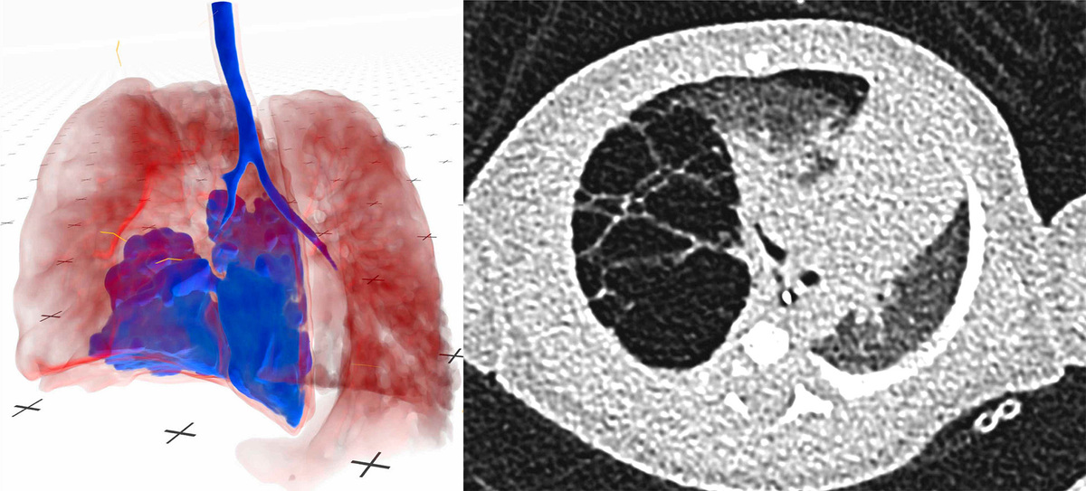
Zalepugas D, Buermann J, Senkel S, Schmidt N, Ziegler AM, Kurz R, Schmidt J, Feodorovici P, Arensmeyer J (2025) The potential of Real-Time volume-rendered 3D Imaging in immersive virtual reality (VR) for surgical planning in infants with congenital thoracic malformation (CTM). Computational and Structural Biotechnology Journal.
The article shows that immersive VR visualization of low-dose CT scans improves surgical planning in pediatric cases with congenital thoracic malformations. Although low-dose CT reduces radiation exposure, it makes anatomy harder to interpret in 2D. Converting these scans into patient-specific 3D VR models gives surgeons a clearer spatial view of organs and malformations than standard CT images.
The results show that VR leads to more accurate diagnoses and more precise surgical decisions, including changes in treatment strategy in several cases. Surgeons identified anatomical details that were missed in 2D imaging, with the strongest benefit seen in less experienced clinicians. The article concludes that VR enables high-quality surgical planning while keeping radiation exposure low.
Patient-specific models
3D visualizations of patient anatomy
in VR are increasingly used across medical fields, including pediatric surgery, to enhance preoperative planning. These tools enable surgeons to view critical structures in axial, sagittal, and coronal planes, improving their understanding of complex anatomy. The integration of AI algorithms streamlines the segmentation process, reducing the time required to identify anomalies and surrounding organs.
Surgeons used 3D Slicer to create a segmented model of an infant's thoracoabdominal organs to identify a cavitary lesion in the right lung lobe. They also applied the tool to visualize an adolescent patient's anatomy, adjusting the opacity of the pancreas and neighboring organs to examine deeper structures around a pseudopapillary tumor. This approach allowed them to plan the dissection with a clearer understanding of the spatial relationships between key structures.
Lung segmentectomy
Researchers in the Netherlands employed a
system combining VR and AI for preoperative planning in congenital lung surgery. A pediatric surgeon and a radiologist first reviewed 2D CT scans of bronchovascular anatomy in five patients, then compared those with corresponding 3D AI-segmented models viewed in VR. While both specialists correctly localized the lesioned lung lobes on conventional imaging, the immersive VR visualizations revealed more affected segments. As a result, in three cases they opted for segmentectomy rather than full lobectomy.
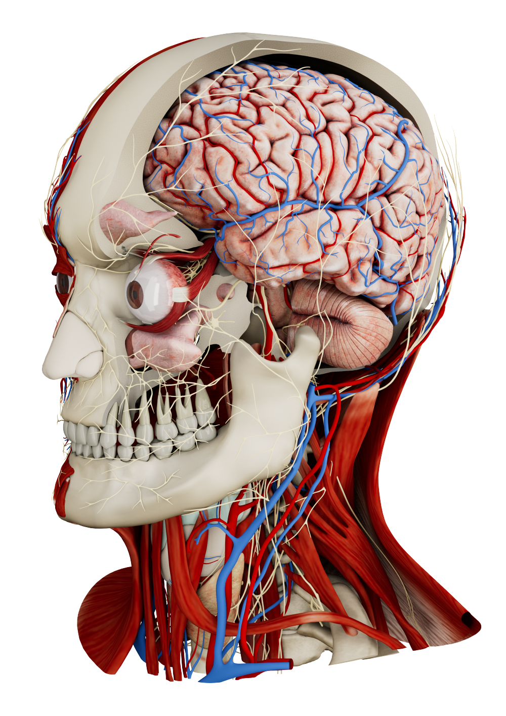
Neurosurgical trajectory planning
Virtual reality is increasingly applied in pediatric neurosurgery to improve comprehension of complex spatial relationships between brain structures. Neurosurgeons applied VR to
optimize surgical trajectories for five children with deep-seated brain lesions near critical vessels and fiber tracts. 2D imaging was converted into 3D models, allowing the surgeons to first plan "top-down" trajectories from the skull surface to the lesion, and then explore "bottom-up" or "canopy" views from beneath or within the lesion to identify alternative or safer paths. These VR fly-throughs enabled them to adjust entry points, avoid critical anatomy, and optimize surgical corridors.
AR-assisted tumor localization
Chest wall tumor resections in pediatric patients often require rib removal, which can result in deformities that impair respiration and mobility. Preserving as much healthy tissue as possible demands precise tumor localization and meticulous surgical technique. Augmented reality technology enabled surgeons to
project patient-specific 3D data onto the pre-incision anatomy
by registering anatomical landmarks and facilitating accurate tumor localization in eight patients. The AR-guided positions were then verified through manual palpation and thoracoscopy. A subsequent cadaver study proved the feasibility of the surface matching method after incision.
Pediatric care is increasingly enhanced by immersive technologies that improve visualization and surgical precision. By integrating 3D medical data, these tools provide a clearer understanding of anatomy and support more informed decision-making. Continued development of these solutions promises safer, more efficient, and patient-centered care for children.
How to get started
Medicalholodeck integrates with secure hospital systems, providing PACS access, HIPAA-compliant data handling, and full patient data security. It runs on stereoscopic 3D displays, VR headsets, mobile devices, and standard Windows systems, enabling flexible use in hospitals, classrooms, and training centers.
Specialized features for surgical planning are exclusive to Medical Imaging XR PRO. Currently, Medicalholodeck is available only for educational use. The platform is undergoing FDA and CE certification, and we expect Medical Imaging XR PRO to be available soon in the U.S. and EU markets.