PRO FDA / CE
Hardware: 3D screen | Virtual Reality i
Runs on 3D and 2D screens on Windows, and Virtual Reality (PC-VR recommended).

Surgical planning

Enhancing surgery with spatial imaging.

Surgical planning is a critical step in preparing for medical procedures. By allowing clinicians to study patient-specific anatomy before entering the operating room, it enhances safety, precision, and overall outcomes. With spatial imaging using virtual reality and stereoscopic 3D screens, traditional CT and MRI scans can be transformed into immersive 3D models, enabling surgeons to explore anatomy in true-to-scale detail and identify the optimal surgical approach with greater confidence.

Important notice regarding surgical planning and professional clinical use
Specialized functionalities for surgical planning and pre-operative professional applications are exclusive to Medical Imaging XR PRO FDA. This version is not yet available. Information about the release date will be published here soon.

Medicalholodeck is currently undergoing the required FDA (U.S. Food and Drug Administration) and CE (Conformité Européenne) certification processes. Our team is working diligently to ensure full compliance with all regulatory standards, and we expect Medical Imaging XR PRO to be available in both the United States and the European Union soon.

For updates on product releases, regulatory progress, and availability, or for any related inquiries, please contact info@medicalholodeck.com.

Risk-free surgical simulations with digital twins

Digital twins are realistic, data-driven 3D replicas of a patient’s anatomy, created from imaging data such as CT or MRI scans. These interactive models mirror real-world structures in precise detail and allow surgeons to scale, rotate, and focus on specific tissues or regions, providing complete control over their visual perspective.

This high level of interactivity enables the simulation of procedures, testing of different surgical approaches, anticipation of potential complications, and enhanced spatial understanding of patient-specific anatomy.

By offering a risk-free environment for professional communication, preoperative training and procedural rehearsal, digital twins help surgical teams refine techniques, optimize outcomes, and improve collaboration and communication.

Case studies in spatial surgical planning

Precision surgery planning in pediatrics

Preoperative planning

Yang W, Xu Y, Wang Z, Ye M, Chen R, Da M, Qi J (2025) Virtual reality-assisted preoperative planning for pediatric thoracoscopic segmentectomy: a retrospective study. BMC Pediatrics.

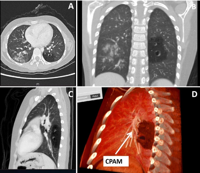
The report describes how pediatric surgeons used Medicalholodeck’s virtual reality medical imaging to support preoperative planning for thoracoscopic lung surgery in children with congenital lung malformations. By transforming CT scans into interactive, patient-specific 3D VR models, surgeons were able to better understand the spatial relationships between lung segments, lesions, and critical vessels than with conventional 2D or standard 3D imaging.

These immersive models were used to plan surgical approaches in advance and to discuss cases within the surgical team. In a series of pediatric thoracoscopic segmentectomies, the VR-based planning helped surgeons navigate complex anatomy more confidently, avoid damage to vital structures, and complete all procedures without conversion to open surgery. Independent senior surgeons confirmed that the VR models provided clearer anatomical insight than traditional imaging alone.

Read more: https://doi.org/10.1186/s12887-025-06259-3

Spatial navigation in mandibular reconstruction

Preprints Image

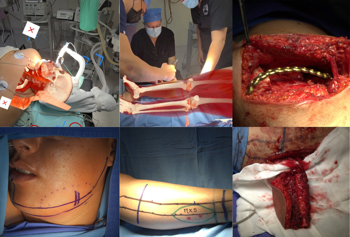
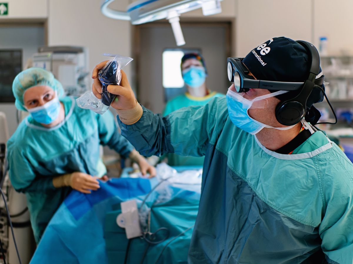
3D holographic navigation for mandibular reconstruction with a free fibula flap. CT-based holograms are overlaid in real time to guide preoperative marking, vascular planning, and microsurgical preparation of donor and recipient vessels.

Mandibular reconstruction using a fibula free flap relies on precise vascular identification, which is technically demanding even with virtual surgical planning. This report describes the first case in Latin America using a low-cost, commercially available augmented reality holographic navigation system to support complex microsurgical reconstruction.

A 26-year-old patient with a large mandibular osteoblastoma underwent preoperative planning using CT-based DICOM data to create 3D holographic models. During surgery, the AR system overlaid these models onto the operative field, enabling real-time identification of displaced recipient vessels in the neck and perforating vessels in the fibula.

An osteofascial fibula free flap was successfully harvested and transferred. The case demonstrates the feasibility and clinical value of affordable, consumer-grade holographic navigation for mandibular reconstruction.

Read more: https://doi.org/10.20944/preprints202508.0103.v1

Orthopedic implant procedures in VR

Virtual reality enables immersive 3D visualization of patient-specific CT data, allowing surgeons to interact with both anatomy and orthopedic implants in a shared virtual environment. Unlike conventional displays, VR provides true depth perception and spatial interaction, enhancing anatomical understanding and implant planning accuracy.

Medicalholodeck, tested by NHS Greater Glasgow and Clyde and NHS Lanarkshire demonstrated proof of concept for preoperative trauma planning using routine imaging and implant files. Using VR hardware and spatial imaging software, the system supports surgical planning and execution, showing high potential to improve fracture comprehension and surgical precision.

Read more: https://doi.org/10.7759/cureus.91198

Spatial telemedicine

XR- and AI-enhanced 3D imaging enable surgeons to collaborate globally in real time. In a study conducted between Uganda and Germany, 3D patient models were created from smartphone photographs and reviewed collaboratively by surgeons in both countries using VR headsets.

The approach was fast, resource-efficient, and allowed precise planning of reconstructive surgeries. It demonstrates how specialists from other countries can provide expert guidance and support to surgeons, even across great distances, improving surgical care and training settings.

Read more: https://doi.org/10.2196/69300

Spatial imaging in spinal surgery

The benefits of virtual reality for surgical planning have been confirmed in multiple studies and real-world cases. This publication presents a 79-year-old patient with thoracic myelopathy and pseudoarthrosis who underwent a two-stage spinal revision.

The procedure was planned using spatial computing in an interactive virtual reality environment, enabling precise optimization of graft size and trajectory. The successful outcome illustrates how immersive 3D planning enhances surgical precision and supports individualized strategies in complex spine surgery.

Read more: https://doi.org/10.4103/jcvjs.jcvjs_48_23

Spatial medical imaging for safer and more efficient procedures

Spatial imaging and virtual reality are transforming surgery by bridging the gap between planning, training, and real-world performance. They allow surgeons to rehearse procedures in a realistic, risk-free environment, improving precision, reducing complications, and building confidence before entering the operating room.

These accessible and cost-effective technologies provide objective performance feedback and unlimited opportunities for practice, accelerating skill development and supporting consistent training standards. They also enable real-time collaboration among specialists worldwide, facilitating expert consultation and shared decision-making.

When combined with AI, VR further enhances surgical planning and execution through improved visualization, streamlined workflows, and increased accuracy. Together, these technologies enable faster preparation, safer procedures, and better patient outcomes.

How to get started

Medicalholodeck integrates with secure hospital systems, providing PACS access, HIPAA-compliant data handling, and full patient data security. It runs on stereoscopic 3D displays, VR headsets, mobile devices, and standard Windows systems, enabling flexible use in hospitals, classrooms, and training centers.

Specialized features for surgical planning are exclusive to Medical Imaging XR PRO. Currently, Medicalholodeck is available only for educational use. The platform is undergoing FDA and CE certification, and we expect Medical Imaging XR PRO to be available soon in the U.S. and EU markets.

For updates, regulatory news, availability, or questions contact info@medicalholodeck.com.

Medical Imaging - Surgical planning and training with digital twins
Pro level
Hardware: Standalone | PC-VR | iOS i
Runs on standalone devices like Meta Quest 3, tethered PC-VR, and iOS devices.

MEDICAL IMAGING

Spatial surgical training.

Learn more Buy
Medicalholodeck AI - Medical imaging and segmentation
Expert level
Hardware: PC-VR | Remote Rendering i
Runs best on tethered PC-VR. Also available on Remote Rendering.

MEDICALHOLODECK AI

Fast automatic segmentation.

Learn more Buy

RecordXR

Record, replay, share.
In augmented and virtual reality.

Learn more Buy

RecordXR Studio

Spatial content editing.

Learn more Buy
Beginner level

ANATOMY MASTER

The spatial anatomy atlas.

Learn more Buy
Anatomy Master Model
Advanced level

DISSECTION MASTER

Real human anatomy.

Learn more Buy
Dissection Master Model